
Nutrition cannot replace oral hygiene, professional biofilm control, or dental treatment. It can, however, influence tissue repair, bone metabolism, immune function, and the ability to maintain healthy tissues around an implant.
This second part examines vitamins, minerals, protein, patient age, medication use, functional capacity, and the need for individualized supportive care.
Authors





Vitamins, minerals, and protein: supporting peri-implant tissues
The long-term maintenance of a dental implant also depends on the body’s capacity for epithelial regeneration, collagen synthesis, bone remodeling, and resolution of inflammation. These processes require an adequate intake of energy, protein, vitamins, and minerals. Nutritional deficiencies are not an independent cause of peri-implantitis, but they may weaken the tissues surrounding an implant and impair healing.
This is particularly important in older adults, patients following elimination diets, people with gastrointestinal disorders, and those taking multiple medications. In these groups, even an apparently varied diet may not ensure adequate absorption of all essential nutrients.
Vitamin C: collagen, blood vessels, and immune function
Vitamin C is one of the best-studied nutrients affecting gingival health. It is involved in the synthesis of collagen, a major component of the connective tissue surrounding an implant. It is also important for normal vascular function, wound healing, and immune-cell activity.
Vitamin C deficiency may weaken gingival structure, increase capillary fragility, delay healing, and increase the tendency to bleed. Reviews indicate that low vitamin C intake or low vitamin C levels are associated with poorer periodontal health. Supplementation may reduce gingival bleeding in people with a deficiency or gingivitis, but there is no conclusive evidence that vitamin C alone prevents advanced bone loss or treats peri-implantitis [8].
The best sources of vitamin C are fresh vegetables and fruit, particularly peppers, parsley, broccoli, cabbage, berries, kiwifruit, and citrus fruit. Regular consumption of these foods is preferable to the occasional use of very high-dose supplements.
It is also important to remember that smoking increases oxidative stress and the body’s requirement for vitamin C. Periodontal disease and poorer outcomes of implant treatment are also more common among smokers. Smoking cessation is therefore far more important than merely increasing vitamin C intake.
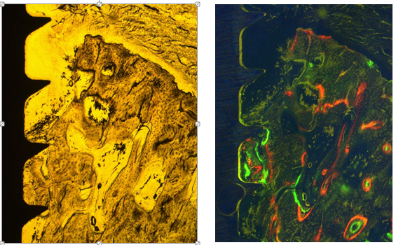
Figure 1. What peri-implantitis means: bacteria-associated bone changes. The upper portion shows bone resorption and loss of bone-to-implant contact. Histology of the coronal part of the implant: undecalcified ground section; A – native image, ×2.5 magnification; fluorescence method, ×2.5 magnification.
Vitamin D3 and calcium: importance for bone health
The bone surrounding an implant is a dynamic structure. Throughout life, it undergoes continuous remodeling involving bone-forming and bone-resorbing cells. Vitamin D3 regulates calcium-phosphate homeostasis, affects bone mineralization, and helps modulate the immune response.
Vitamin D3 deficiency may impair bone healing after implant placement. Studies suggest an association between low vitamin D3 levels and poorer osseointegration or a higher incidence of early implant failure. In addition to modulating bone formation, vitamin D affects innate and adaptive immune responses within osteoimmunology and may therefore influence early implant healing by: 1) upregulating the expression of genes encoding osteocalcin, osteopontin, calbindin, and 24-hydroxylase; 2) increasing extracellular-matrix protein production by osteoblasts; and 3) stimulating osteoclast activity. Vitamin D deficiency may therefore disrupt the delicate balance between the immune system and bone metabolism during implant healing through direct or indirect changes in osteoclast function.
However, the quality of the available evidence remains variable, and most studies concern initial implant osseointegration rather than the direct development of late peri-implantitis caused by vitamin D deficiency itself [9].
It should nevertheless be emphasized that vitamin D works together with cofactors, including vitamins K2, A, and C and magnesium, in processes that determine its biological activity. Through its pleiotropic effects and multiple hormonal pathways, vitamin D may indirectly regulate immune function and thereby influence the potential development of early or late peri-implantitis. The supplementation dose and duration are also important, and this is where controversy arises. The Institute of Medicine (IOM) issues dietary recommendations at the request of the governments of the United States and Canada. One such recommendation is the Recommended Dietary Allowance (RDA), defined as an intake sufficient to meet the nutrient requirements of 97.5% of healthy individuals [17].
The RDA for vitamin D is 600 IU per day for people aged 1-70 years and was assumed to achieve a serum 25-hydroxyvitamin D [25(OH)D] concentration of at least 50 nmol/L in 97.5% of healthy individuals [1]. Serum 25(OH)D is the established indicator of vitamin D status, and concentrations of at least 50 nmol/L have been associated with bone health and protection against disease and injury [1]. New analyses using linear regression reported that 600 IU of vitamin D per day would result in 97.5% of individuals having serum 25(OH)D concentrations above 26.8 nmol/L rather than above the currently assumed 50 nmol/L threshold. It was also estimated that 8,895 IU of vitamin D per day might be required for 97.5% of individuals to reach serum 25(OH)D concentrations of at least 50 nmol/L. Because this dose substantially exceeds the range of doses studied, the estimate should be interpreted with caution. Nevertheless, the high estimate illustrates that the calculated dose greatly exceeds both the current RDA of 600 IU/day and the tolerable upper intake level of 4,000 IU/day [1].
It should therefore not be claimed that vitamin D3 supplementation alone prevents peri-implantitis. In patients with a confirmed deficiency, correcting the deficiency may support bone metabolism, immune and endocrine function, and overall health. Older adults, patients with osteoporosis, people who avoid sun exposure, patients with malabsorption disorders, and those undergoing cancer treatment require particular attention, including those at risk of osteonecrosis of the jaw, whether medication-related or radiation-induced [18-23].
Vitamin K2
Two natural forms of vitamin K occur in nature: vitamins K1 and K2. Both share a 2-methyl-1,4-naphthoquinone ring but differ in the type and length of the isoprenoid side chains attached to it. Vitamin K1, also known as phylloquinone or phytomenadione, has a phytyl chain consisting of one unsaturated and three saturated isoprenoid units. Vitamin K2 compounds, known as menaquinones and designated MK followed by a number indicating the number of isoprenoid units, contain side chains composed exclusively of 4-10 unsaturated isoprenoid units. Vitamin K is sensitive to acidic and alkaline conditions and to light.
Vitamin K is absorbed from the intestine into the lymphatic system. Normal absorption requires bile and pancreatic juice. Absorption varies by form: vitamin K1 requires energy and the presence of long-chain unsaturated fatty acids, whereas vitamin K2 (menaquinone) and vitamin K3 are absorbed by diffusion.
Vitamin K intake is associated with changes in calcium homeostasis and may promote calcium deposition in bone. The active form of vitamin D3, 1,25-dihydroxycholecalciferol (calcitriol), together with vitamin K, is important for osteocalcin production by osteoblasts. Osteocalcin is a low-molecular-weight protein produced by osteoblasts, odontoblasts, and chondrocytes. Its blood concentration is a marker of osteoblast activity. It participates in bone mineralization and calcium regulation and acts as a tissue hormone. Most importantly, vitamin K is essential for blood coagulation because it enables the carboxylation of glutamic acid residues in seven proteins involved in coagulation.
Vitamin K is rapidly excreted in urine or feces. Its half-life is only 72 hours.
Vitamin K2 MK-7 deficiency may occur in newborns because they lack vitamin-producing microbiota and breast milk contains only small amounts, and in older adults because of gastrointestinal dysfunction and the use of antibiotics or other medicines. Vitamin K deficiency affects blood coagulation and bone density, manifesting as a tendency to bleed, impaired bone mineralization, and increased vascular calcification.
Calcium is the principal mineral component of bone, but its effects are closely linked to vitamin D3. Taking large amounts of calcium without assessing the diet, vitamin D3 status, and general health does not always provide measurable benefits. Natural calcium sources include milk and fermented dairy products, fish eaten with their bones, certain leafy vegetables, almonds, and fortified foods.
The decision to supplement vitamin D3 or calcium should take account of laboratory results, comorbidities, and current medication. This is especially important in patients with kidney disease, disorders of calcium metabolism, or nephrolithiasis.
Boron is a trace element that supports calcium and magnesium homeostasis and may affect vitamin D3 metabolism and persistence in the body. Although present only in low concentrations in bone, muscle, and the brain, it performs important biological functions.
Boron influences calcium and magnesium metabolism and calcium homeostasis, vitamin D absorption and maintenance of its concentration, the synthesis of hormones such as testosterone and estrogen, wound healing and tissue repair, and the modulation of inflammation, particularly in the joints.
Boron occurs mainly in plant foods. The richest sources include leafy vegetables, stone fruit, legumes, and nuts.
Magnesium and vitamin D
All enzymes that metabolize vitamin D require magnesium, which acts as a
COFACTOR in reactions occurring in the liver and kidneys
Role of magnesium: magnesium activates not only vitamin D but also more than 600 enzymes in the body. It also affects extracellular calcium concentrations and, by forming complexes with phospholipids, is an integral component of cell, intracellular-organelle, mitochondrial, and endoplasmic-reticulum membranes. It also stabilizes ribosomes, lysosomes, and mitochondria. Magnesium is necessary for maintaining stable cellular function, RNA and DNA synthesis, cell repair, and cellular antioxidant status. Approximately 40% of the body’s magnesium is intracellular, and almost 60% is found in bones and teeth; less than 1% is present in extracellular fluids, including serum. Serum magnesium concentration therefore does not reflect total body magnesium status. A blood test may be normal even when intracellular magnesium content is markedly low and symptoms typical of deficiency persist [24].
Vitamins A and E: epithelial protection and antioxidant activity
Vitamin A participates in the differentiation and renewal of epithelial cells. Deficiency may impair mucosal health and weaken its protective function. This does not mean, however, that additional vitamin A will improve peri-implant tissue health in a person without a deficiency. Vitamin A can accumulate in the body; therefore, high-dose supplementation should be used only after clinical indications have been assessed.
Vitamin E is part of the antioxidant system that protects cell membranes from damage. Studies of periodontal diseases have evaluated vitamin E together with vitamin C, selenium, and other antioxidants. Improvements in selected clinical parameters have sometimes been reported, but current findings do not support recommending such preparations for the routine treatment of peri-implantitis.
A varied diet containing vegetables, fruit, nuts, seeds, and high-quality plant oils remains a safer source of antioxidants and the fat-soluble vitamins A, D, E, and K. Figures 2-5 below show the vitamin A, D, E, and K content per 100 g of food (materials courtesy of dietitian Arkadiusz Matras and reproduced with permission).
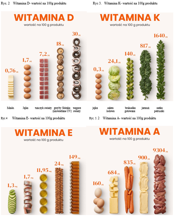
Figures 2–5. Vitamin D, K, E and A content per 100 g of selected foods.
Figures 6-8 present reference ranges for vitamin D and its supplementation with cofactors. It is very important to perform a follow-up test three months after starting appropriate supplementation prescribed by a physician on the basis of the current vitamin D concentration measured in capillary or venous blood. Depending on the result, supplementation may need to be continued until an appropriate, stable vitamin D level has been achieved.
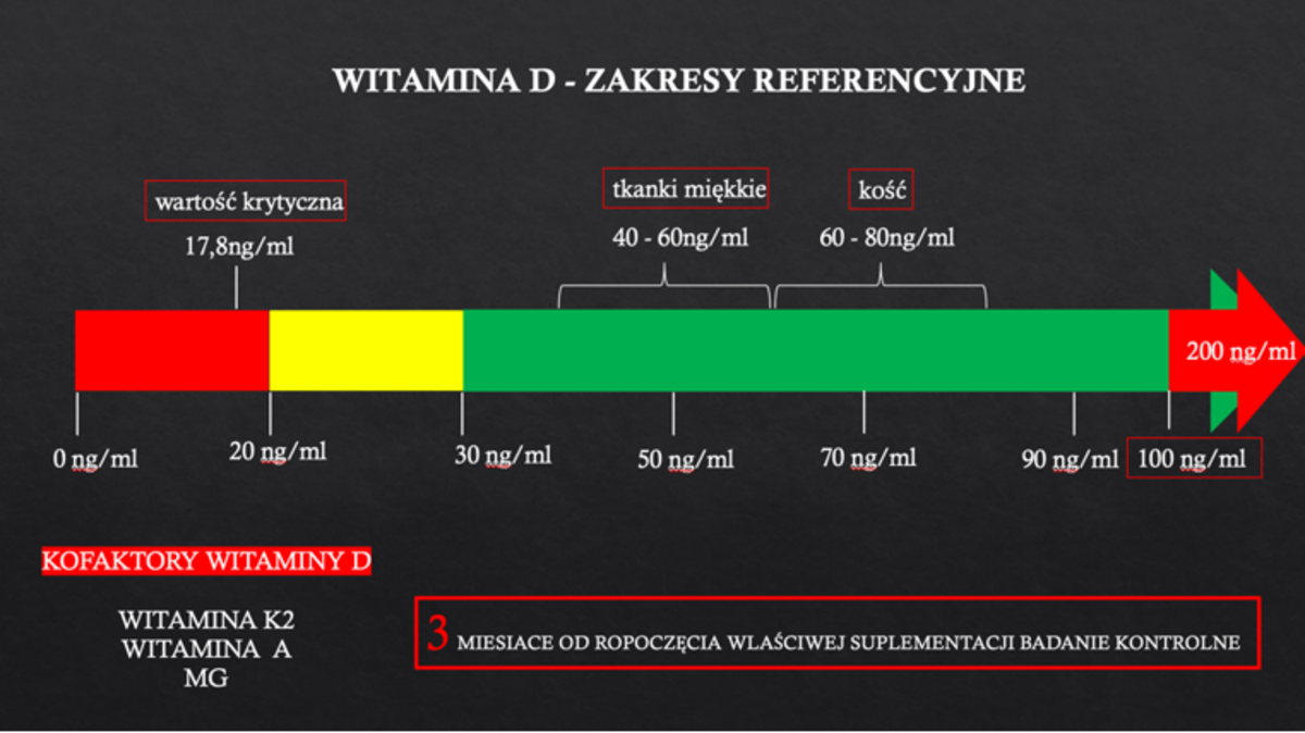
Figure 6. Vitamin D reference ranges (adapted from Marzena Dominiak, “Oral Health Prevention,” in Health Education: A Textbook for Teachers, scientific editors Jolanta Grzebieluch, Alicja Basiak-Rasała, and Barbara Grabowska, Wroclaw Medical University, Wroclaw, 2025).
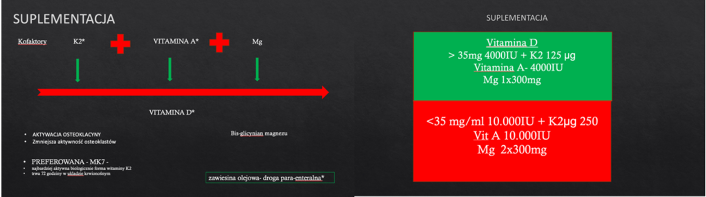
Figures 7 and 8. Principles of vitamin D supplementation with cofactors and supplementation doses according to laboratory test results.
Vitamin B12 and folate: particularly important in older age
B-group vitamins participate in cell division, hematopoiesis, and tissue regeneration. Vitamin B12 deficiency may cause anemia, weakness, a burning sensation of the tongue, oral mucosal lesions, and neurological disorders. Folate plays an important role in cell division and is therefore essential for normal tissue regeneration, including epithelial-cell renewal.
Evidence regarding a direct association between vitamin B12 deficiency and peri-implantitis remains insufficient. B12 deficiency should therefore not be regarded as a confirmed cause of this disease. Periodontal studies nevertheless indicate that inadequate intake of B-group vitamins, especially folate and vitamin B12, may coexist with poorer periodontal health.
The risk of vitamin B12 deficiency increases in older adults, patients with gastrointestinal disease, people who have undergone bariatric surgery, and those taking certain medicines over a long period, including metformin or gastric acid-suppressing drugs. Vegans are also at risk because natural sources of vitamin B12 are primarily foods of animal origin. Periodic laboratory assessment and appropriately selected supplementation should therefore be considered in these individuals.
Protein: an essential substrate for tissue regeneration
The soft tissues surrounding an implant undergo continuous renewal. Protein is required for the synthesis of collagen, enzymes, antibodies, and mediators involved in healing. Deficiency may weaken immune function, reduce muscle mass, and delay wound healing.
In experimental studies, a low-protein diet impaired the integration of titanium implants with bone. These findings cannot be directly extrapolated to routine clinical practice or used to regard protein deficiency as an independent cause of peri-implantitis. They do, however, indicate that adequate nutritional status is an important prerequisite for healing and tissue maintenance [10].
Protein deficiency is particularly relevant to malnourished individuals, older adults, patients after extensive surgery, people with chronic diseases, and patients who have difficulty chewing or swallowing. Good protein sources include fish, eggs, dairy products, lean meat, legumes, and soy products.
Diet first, supplementation second
Supplements do not remove bacterial biofilm and cannot replace dental treatment. Their use is best justified in cases of a confirmed deficiency, increased requirement, or limited ability to obtain a nutrient from the diet.
In a patient with recurrent inflammation, impaired healing, or progressive bone loss, assessment of general health and dietary intake should be considered. Depending on the clinical situation, the clinician may request tests including vitamin D status, a complete blood count, and assessment of vitamin B12, folate, iron, or parameters of calcium and lipid metabolism.
Supplementation should primarily be used to correct confirmed deficiencies and cannot replace a properly balanced diet. A varied diet providing adequate protein, vegetables, fruit, whole-grain products, fish, and fermented dairy products remains the foundation of prevention and maintenance of peri-implant tissue health.
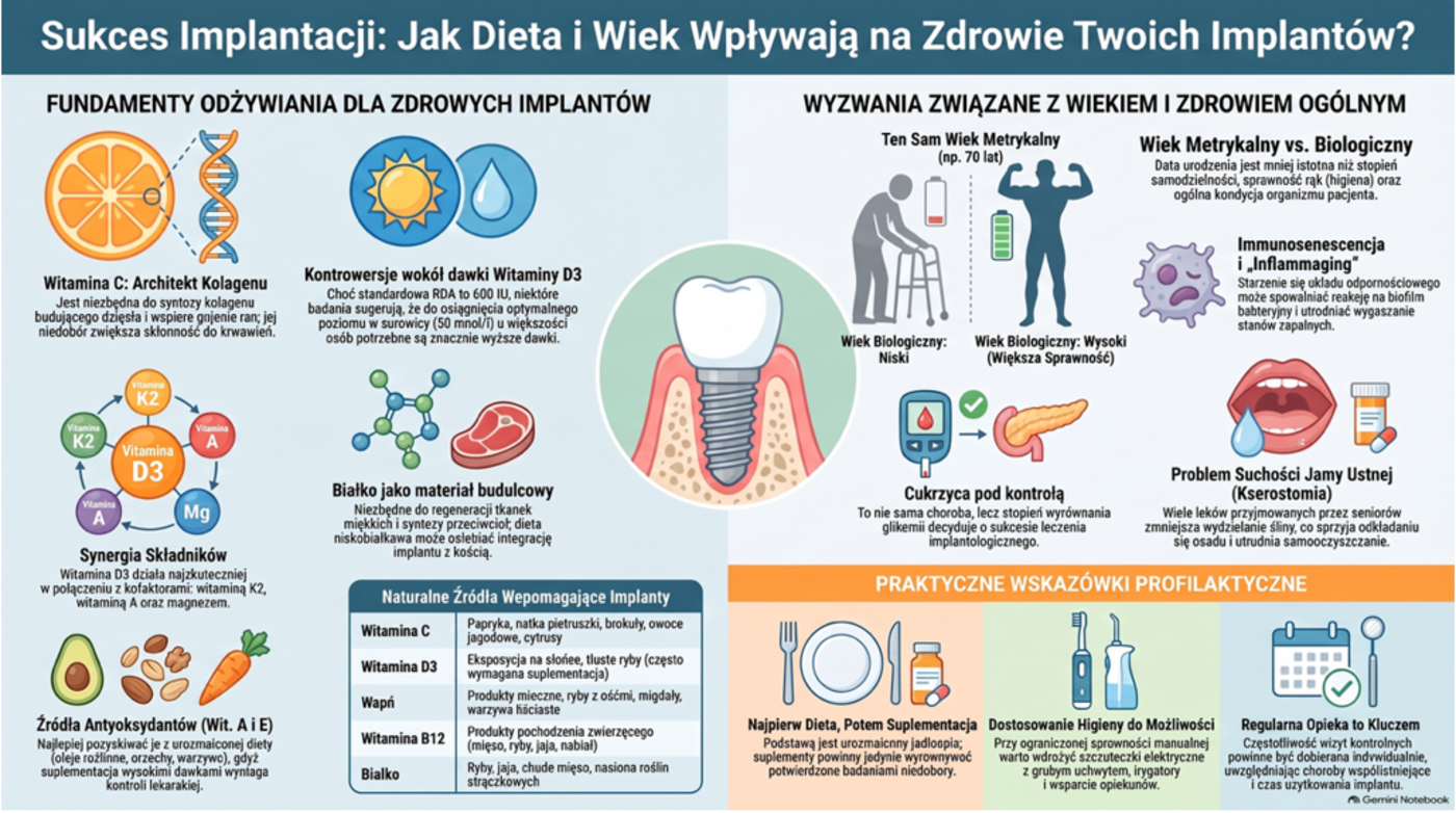
Figure 9. Effects of diet and patient age on the risk and course of peri-implantitis.
Patient age and the risk of peri-implantitis
Dental implants are increasingly used in older adults because they improve chewing ability, eating comfort, and quality of life. Well-planned treatment can provide very good outcomes even in patients in their seventies or eighties. Advanced age alone is neither a contraindication to implant placement nor a clearly established independent risk factor for peri-implantitis. General health, history of periodontitis, control of metabolic diseases, smoking, functional capacity, and the ability to maintain adequate oral hygiene are more important than chronological age [4]. Recent reviews emphasize that chronological age should not automatically be equated with a greater risk of implant-treatment failure.
Chronological age is not the same as biological age
Two people aged 75 years may differ considerably in their health status. One may remain active, eat well, maintain oral hygiene independently, and attend regular follow-up visits. The other may have several chronic diseases, take multiple medications, have limited manual dexterity, memory impairment, and malnutrition. The risk of peri-implantitis should therefore be assessed on the basis of biological age and the patient’s degree of independence, not merely date of birth.
Diseases and disorders that may indirectly affect peri-implant tissues are more common in older adults. These include diabetes mellitus, osteoporosis, cardiovascular disease, chronic kidney disease, anemia, malabsorption disorders, and impaired immune function. A history of periodontitis is also important and is one of the best-documented factors associated with an increased risk of peri-implantitis.
Ageing of the immune system
Ageing is a time-dependent process whose rate is influenced by both genetic and environmental factors. Its consequences include a progressive loss of tissue and organ integrity and impaired function, which contribute to chronic disease. In 1969, Walford proposed the “immunologic theory of ageing,” which subsequently evolved into the concept of immunosenescence, encompassing the changes that occur in the immune system with age. Immunosenescence does not mean a complete loss of immunity but rather a reduced ability to respond rapidly and precisely to microorganisms. At the same time, some older adults develop persistent low-grade inflammation, often termed “inflammaging.” Immunosenescence and inflammaging are therefore regarded as two pillars of age-related immune dysfunction.
| IMMUNOSENESCENCE | INFLAMMAGING |
|---|---|
| Age-related decline in the function of immune-system organs (thymus, spleen, skin) | Chronic low-grade inflammation |
| Impaired physical and mucosal barrier function | Accumulation of senescent cells with a senescence-associated secretory phenotype (SASP) |
| Reduced hematopoietic stem-cell (HSC) activity | Metabolic disturbances and mitochondrial dysfunction |
| Reduced innate and adaptive immune responses | Gut dysbiosis and microbial imbalance |
In the PERI-EDU consortium’s own study, a multivariable model adjusted for age, sex, osteoporosis, hypertension, and cardiovascular disease showed that diabetes was associated with the presence of focal low-density intramedullary trabecular alterations (FLDIFs) (adjusted odds ratio, 1.647; 95% CI, 1.022-2.686; p = 0.042). This estimate should be considered hypothesis-generating because information on systemic diseases was collected retrospectively, important confounders were unavailable, and sensitivity analysis showed that the result was not fully robust. FLDIF is a descriptive CBCT phenotype, not a diagnosis of fatty degenerative osteonecrosis of the jaw, bone-marrow adiposity, focal osteoporotic bone-marrow defect, or another specific disease. CBCT depicts mineralized trabecular architecture and relative attenuation, but it cannot directly demonstrate marrow fat or confirm osteonecrosis.
Nevertheless, inflammation associated with the aforementioned diseases, together with radiographically visible changes and ageing bone marrow, may impair hematopoietic stem-cell (HSC) function. HSC ageing leads to changes in both the number and function of these cells.
Features of immune-cell ageing include impaired immune function, loss of functional capacity, and increased release of proinflammatory mediators.
Under these conditions, the response to bacterial biofilm may be less effective and resolution of inflammation may be slower. Tissues may also require more time to regenerate after a procedure or professional debridement. This does not mean that peri-implantitis is an inevitable consequence of ageing. In an appropriately treated older patient who attends regular follow-up visits, implants can remain stable for many years.
Available evidence suggests that the higher prevalence of periodontal and peri-implant diseases observed in older age groups may result not only from immune changes, but also from longer exposure to biofilm, a greater burden of chronic disease, and a declining ability to maintain oral hygiene independently [4].
An implant also has an “age”
When considering age, the patient’s age must be distinguished from the time the implant has been in function. An implant that has been present in the oral cavity for 15 or 20 years has been exposed to biofilm and recurrent inflammatory episodes for far longer than one placed a year ago.
The greater frequency of peri-implantitis in older adults may therefore be partly attributable to the longer functional period of the prosthetic restoration and to possible consequences of metal corrosion.
No metal or alloy is completely inert in vivo. Because a metallic implant is in contact with tissues and body fluids, ions and particles may be generated and released into the biological environment by:
-
electrochemical processes (corrosion);
-
wear or friction (tribological processes).
When corrosion occurs together with tribological processes, the resulting phenomenon is known as TRIBOCORROSION. When tribocorrosion occurs in a biological system, it is termed BIOTRIBOCORROSION.
Three types of corrosion are most commonly encountered in implant dentistry: 1) galvanic corrosion; 2) fretting corrosion caused by micromovement between contacting surfaces at implant connections; and 3) pitting corrosion occurring on the implant surface in contact with the environment and promoted, for example, by fluoride or bleaching agents. Corrosion particles and wear debris in peri-implant tissues may ultimately trigger a cascade of events leading to peri-implant bone loss. A reduced pH in the electrolytic environment during local inflammation appears to promote corrosion. Whether inflammation and the associated pH reduction cause corrosion, or corrosion initiates an inflammatory response, remains controversial.
Moreover, titanium hypersensitivity or intolerance in dental and maxillofacial implant surgery may be more common than reported in the literature. Such a reaction to titanium could account for otherwise unexplained repeated dental-implant failures in some patients. Wroclaw Medical University has therefore initiated a prospective, exploratory clinical study within the international PERI-EDU consortium. The study combines clinical examination, CBCT-based assessment of bone loss, paired blood sampling, and SEM/EDX analysis of explanted implants to investigate how severe peri-implant inflammation may link local tissue destruction with systemic inflammation and corrosion-related surface changes (www.peri-edu.umw.edu.pl).
Over time, cleaning an implant-supported crown, bridge, or denture may also become more difficult. Changes in gingival position, wear of prosthetic components, and exposure of implant surfaces can create plaque-retentive sites and may also contribute to implant corrosion.
The ability to clean the prosthetic restoration thoroughly is also important. Even an esthetic and durable restoration may increase the risk of inflammation if the patient cannot reach the surfaces beneath a bridge or denture with a toothbrush or appropriate cleaning aids.
Manual dexterity, vision, and memory
One of the most important challenges of older age is a declining ability to clean the oral cavity independently. Osteoarthritis, hand tremor, previous stroke, Parkinson’s disease, or muscle weakness may make the precise use of a toothbrush, dental floss, or interdental brushes difficult. Poorer vision limits the ability to notice plaque and bleeding, while memory impairment may lead to missed daily hygiene procedures.
In this situation, simply reminding the patient to brush more often is not sufficient. The hygiene method may need to be adapted to the patient’s actual abilities. Electric toothbrushes with thicker handles, oral irrigators, appropriately sized interdental brushes, and instruction provided together with a caregiver can be helpful.
In patients who are partly or fully dependent on others, implant hygiene should be incorporated into daily care. Loss of independence is a point at which oral health can deteriorate very rapidly, even when implants have functioned well for many years [4].
Dry mouth and medication use
Older adults are more likely to take several or even more than ten medicines concurrently. Many of these can reduce salivary secretion, including certain antidepressants, sedatives, antihistamines, diuretics, and antihypertensive drugs.
Saliva moistens the oral mucosa, facilitates oral clearance, and contains components involved in antimicrobial protection. Reduced salivary flow may cause burning, difficulty chewing and swallowing, halitosis, and increased plaque accumulation. Dry mouth alone is not a sufficient cause of peri-implantitis, but it can make it more difficult to maintain a healthy oral environment.
A patient with persistent dry mouth should inform their dentist. Medicines should not be discontinued without medical advice, but changing the time of administration, having the treating physician adjust therapy, using moisturizing products, and sipping water more frequently may be considered. Sugar-sweetened drinks and sweets used to stimulate salivary flow may do more harm than good.
Age, malnutrition, and vitamin deficiencies
The risk of malnutrition increases with age. Causes may include reduced appetite, loneliness, financial constraints, chronic disease, chewing difficulties, altered taste, depression, and difficulty preparing meals. A patient may consume enough calories while still receiving too little protein, vitamins, and minerals.
Vitamin D3, vitamin B12, vitamin C, calcium, and protein require particular attention. Vitamin D3 deficiency is common in older adults because of limited sunlight exposure and the skin’s reduced capacity for synthesis. Vitamin B12 absorption may also decline with age and with the use of certain medicines.
Inadequate protein intake contributes to loss of muscle mass, or sarcopenia, and may limit tissue-repair capacity. Weaker muscles and declining function further impair the ability to prepare meals and maintain oral hygiene. This creates a vicious cycle linking malnutrition, frailty, and deteriorating oral health.
This does not mean that every older patient with an implant should take a combination of supplements. It is preferable to identify the actual problem, assess the diet, and, when indicated, perform laboratory tests. Supplementation with vitamin D3 and cofactors, vitamin B12, calcium, or iron should be tailored to the patient’s health status because excessive intake of some nutrients can also be harmful.
Diabetes and glycemic control
Diabetes is more common in older age groups and may increase susceptibility to infection and impair healing. More than three million people in Poland have diabetes, and this number is steadily increasing. However, the degree of glycemic control is more important than the diagnosis alone. Chronic hyperglycemia promotes the formation of advanced glycation end products, increases oxidative stress, and may lead to an exaggerated inflammatory response (see above).
Well-controlled diabetes does not preclude successful implant treatment. The patient should nevertheless remain under the care of a diabetologist, monitor blood glucose regularly, and avoid a diet rich in simple sugars and highly processed carbohydrates. Prevention of peri-implantitis includes not only oral hygiene but also metabolic control.
Regular care matters more than chronological age
The greatest threat to an older patient may not be age itself but a loss of continuity of care. Moving home, hospitalization, admission to a care facility, or declining functional capacity may interrupt regular follow-up visits. Lack of supportive care and inadequate hygiene are important factors associated with peri-implant disease.
The recall interval should be individualized. A patient with a history of periodontitis, diabetes, limited dexterity, or a prosthetic restoration that is difficult to clean may require more frequent visits than a low-risk patient. Follow-up should assess not only implant mobility, but also bleeding, probing depth, suppuration, bone levels, and whether the restoration can be cleaned effectively.
Age should therefore not be regarded as a sentence for an implant. It should instead prompt a more individualized approach that considers general health, nutritional status, medication use, independence, and caregiver support. Regular preventive care and prompt management of the first signs of inflammation are more likely to determine treatment success than the patient’s age alone.
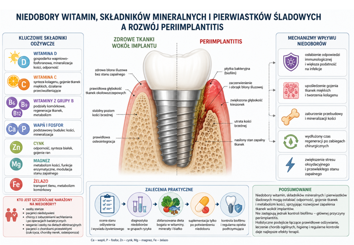
Figure 10. Summary of nutritional factors – vitamins, minerals, and trace elements – that may influence the development of peri-implantitis.
The article was originally published in Polish in Medium, the magazine of the Lower Silesian Chamber of Physicians, October 2026.
Information
Read Part 1: The Impact of Nutrition on Peri-Implantitis.
Follow the training modules: PERI-EDU Module 1 – Peri-implantitis: From Biofilm to Bone Loss.
This material was prepared as part of the “Integrating Peri-Implantitis Research into Higher Education Curriculum” (PERI-EDU) project, co-funded by the European Union under grant agreement No. 2024-1-PL01-KA220-HED-000251208.
Disclaimer
Co-funded by the European Union. The views and opinions expressed are, however, those of the author(s) only and do not necessarily reflect those of the European Union or the Erasmus+ National Agency (NA). Neither the European Union nor the NA can be held responsible for them.