
Osteolytic Jaw Lesions on CBCT: Frequency and Systemic Associations was presented as part of the PERI-EDU research programme during the 9th Congress of the Polish Dental Association at Ptak Warsaw Expo.
Research question
Study design
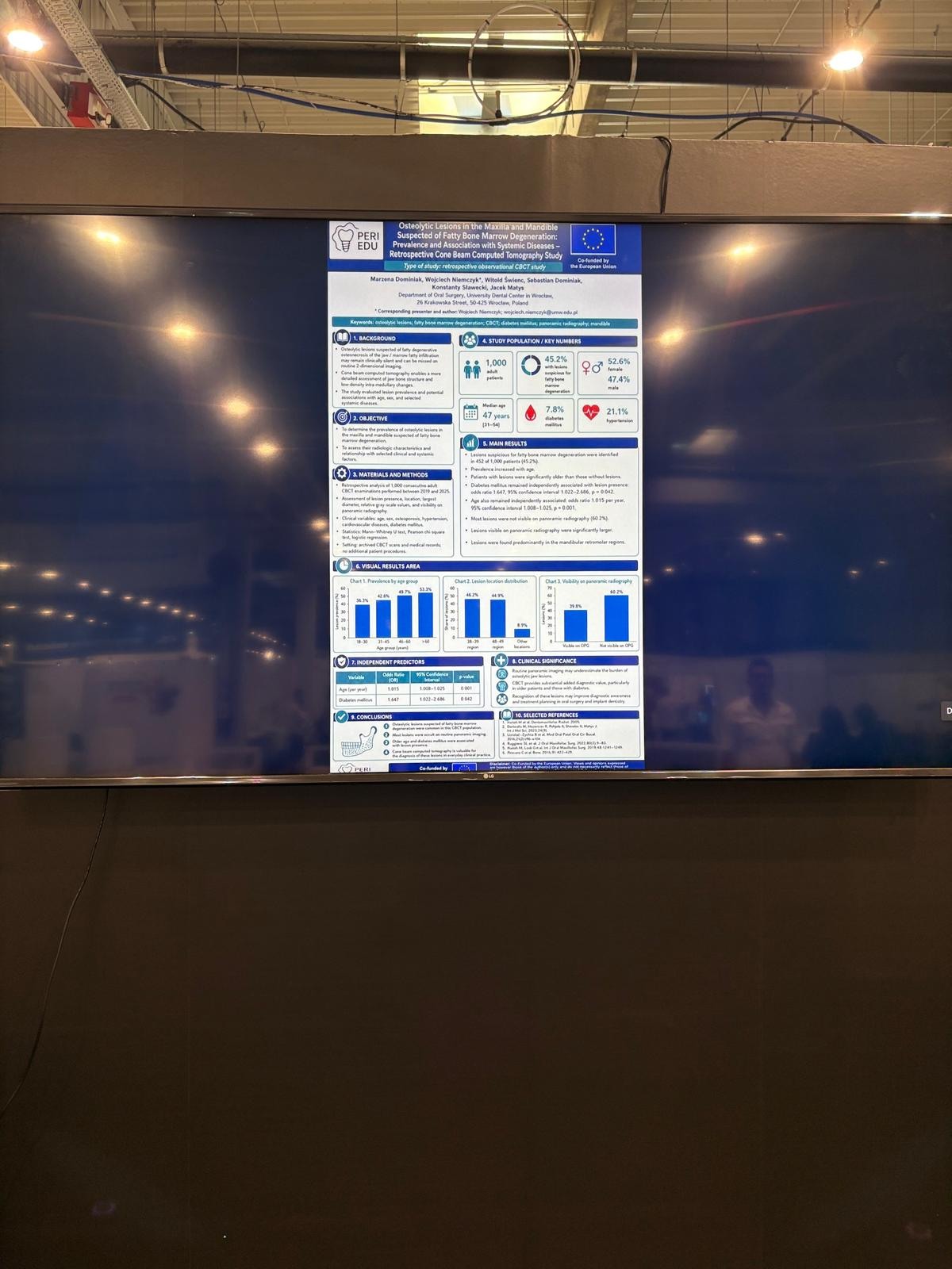
The investigators retrospectively assessed 1,000 consecutive adult CBCT examinations performed between 2019 and 2025. Lesion presence, location, diameter, grey-scale characteristics and panoramic visibility were evaluated together with age, sex and selected systemic diseases.
Main findings
- Suspicious lesions were found in 452 of 1,000 patients (45.2%), predominantly in mandibular retromolar regions.
- Most lesions were not visible on panoramic radiographs (60.2%); lesions visible on panoramic imaging were significantly larger.
- Age remained independently associated with lesion presence (OR 1.015 per year; 95% CI 1.008-1.025; p=0.001).
- Diabetes mellitus was also independently associated (OR 1.647; 95% CI 1.022-2.686; p=0.042).
Interpretation
The findings suggest that routine panoramic imaging may underestimate these low-density jaw-bone changes. The retrospective design identifies associations rather than causation; radiographic findings require careful differential diagnosis and clinical correlation.
Download the full conference poster
The complete poster is available as a PDF, including figures, numerical results and the selected reference list.
Download poster PDF
View congress report
View abstract supplement
Funding: This work was co-funded by the European Union under grant agreement No. 2024-1-PL01-KA220-HED-000251208.
Disclaimer: Co-funded by the European Union. Views and opinions expressed are, however, those of the author(s) only and do not necessarily reflect those of the European Union or the Foundation for the Development of the Education System (FRSE). Neither the European Union nor FRSE can be held responsible for them.