
Focal Low-Trabeculation and Low-Density Jawbone Findings on CBCT: Frequency and Systemic Associations
A retrospective study of 1,000 adults examined how often a study-defined, nonspecific intramedullary jawbone imaging phenotype was identified on CBCT and explored its relationships with age, systemic diseases, anatomical location and visibility on panoramic radiographs.
Why was the study performed?
Focal areas of reduced cancellous trabeculation and relatively low radiographic density may be encountered on dental CBCT, especially in the posterior mandible. Their interpretation is difficult because similar appearances may reflect physiological or age-related marrow variation, focal fatty replacement, focal osteoporotic bone marrow defect or other local processes.
The study aimed to determine the frequency of a strictly defined radiological phenotype within a selected cohort referred for CBCT and to explore its associations with age, sex, osteoporosis, arterial hypertension, cardiovascular disease and diabetes mellitus. It also assessed the anatomical distribution and radiographic characteristics of the largest index finding in each positive patient and whether the same site was appreciable on panoramic radiography.
Study design
Adults
Archived CBCT examinations meeting predefined image-quality and anatomical-coverage requirements.
Positive patients
At least one study-defined FLDIF was identified in 452 patients.
Cohort frequency
A cohort-specific proportion, not an estimate of prevalence in the general population.
Index findings
One largest finding per positive patient was used for finding-level analyses.
The examinations had been acquired previously for routine clinical reasons; no CBCT scan was performed specifically for the research. The patient was the primary unit of analysis. If several findings were present, only the largest was selected as the index finding, avoiding correlated measurements but favoring larger lesions in the finding-level description.
Key findings
1. Frequency increased across age groups
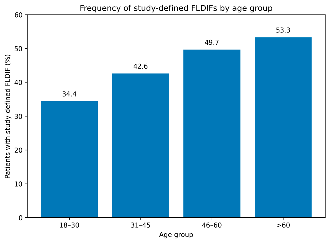
Study-defined FLDIFs were identified in 452 of 1,000 patients (45.2%; 95% CI: 42.1-48.3%). The observed proportion increased from 34.4% in patients aged 18-30 years to 53.3% in those older than 60 years. In the adjusted model, age was associated with finding presence (OR 1.015 per year; 95% CI: 1.006-1.025), equivalent to an estimated OR of 1.161 per ten-year increase under the model assumptions.
2. Diabetes showed a modest exploratory association
In the multivariable model adjusted for age, sex, osteoporosis, arterial hypertension and cardiovascular disease, diabetes mellitus was associated with FLDIF presence (adjusted OR 1.647; 95% CI: 1.022-2.686; p = 0.042). This estimate should be treated as hypothesis-generating because systemic disease information was obtained retrospectively, important confounders were unavailable and sensitivity analysis showed that the result was not fully stable.
| Variable | Adjusted odds ratio | 95% confidence interval | Interpretation |
|---|---|---|---|
| Age, per year | 1.015 | 1.006-1.025 | Statistical association; physiological marrow conversion remains a possible explanation. |
| Diabetes mellitus | 1.647 | 1.022-2.686 | Modest exploratory association requiring independent confirmation. |
| Sex | 0.927 | 0.719-1.194 | No statistically significant adjusted association. |
| Osteoporosis | 0.743 | 0.292-1.893 | Imprecise estimate; only 20 patients had osteoporosis. |
| Arterial hypertension | 1.140 | 0.810-1.604 | No statistically significant adjusted association. |
| Cardiovascular disease | 1.050 | 0.662-1.665 | No statistically significant adjusted association. |
3. Most index findings were in mandibular retromolar regions
Of the 452 largest index findings, 412 (91.2%) were located in mandibular retromolar regions: 209 on the left and 203 on the right. This describes only the location of the single largest finding per positive patient, not the distribution of every finding present in the cohort.
4. Most CBCT-defined sites were not visible on panoramic radiographs
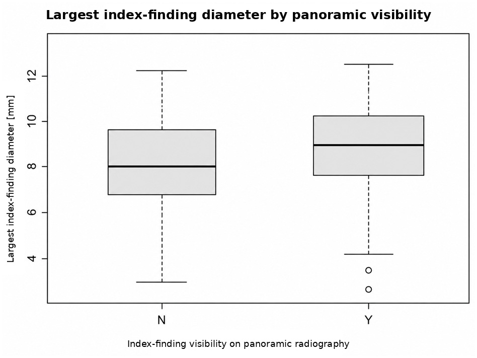
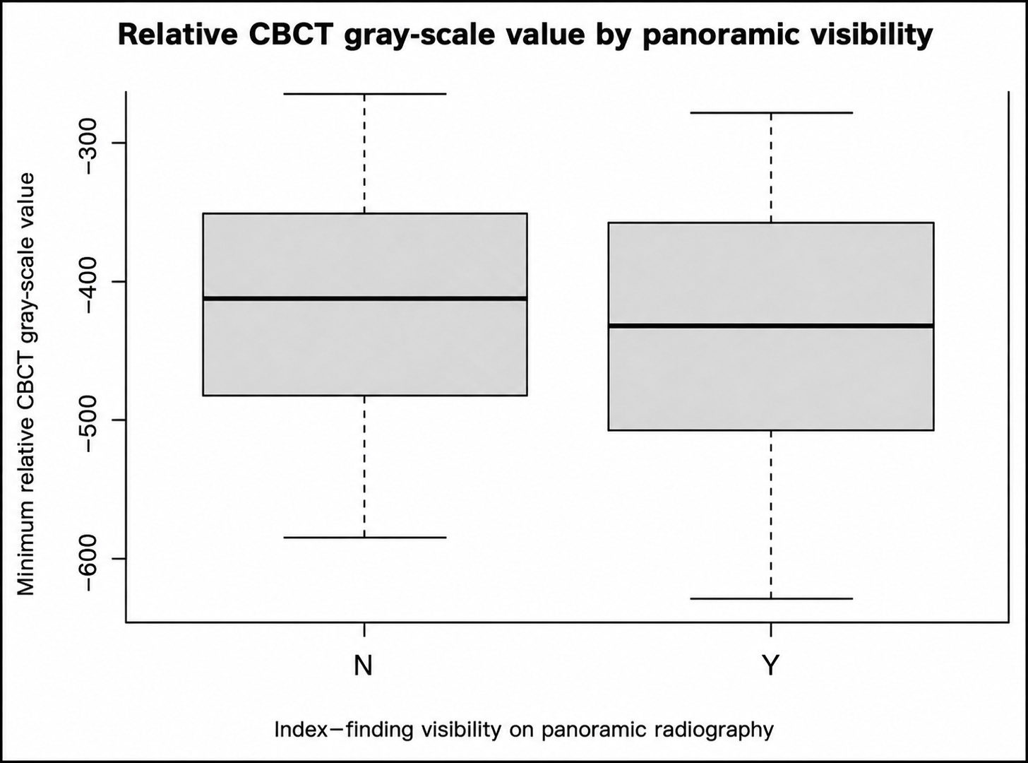
All 452 index findings had a corresponding panoramic radiograph available. Only 180 (39.8%) were judged visible, while 272 (60.2%) were not visible. Findings visible on panoramic radiography were slightly larger than non-visible findings: median diameter 9.2 mm versus 8.1 mm. The effect size was small, and the comparison was targeted to previously CBCT-defined sites rather than designed as a diagnostic-accuracy study.
What do these results mean?
The study identifies a reproducible imaging phenotype that may be useful for future radiological research. It does not determine the pathological nature of the observed areas and does not demonstrate that identifying them changes treatment or patient outcomes. The reported 45.2% is specific to a selected university dental-center cohort already referred for CBCT and must not be generalized to the population.
The study also does not establish a reason to perform CBCT screening based on age or diabetes. CBCT examinations must continue to be justified by accepted clinical indications and radiation-protection principles. When a finding has potential clinical relevance, interpretation should incorporate symptoms, dental and extraction history, clinical examination and, where clinically indicated, complementary imaging or histopathological correlation.
Limitations and next steps
The retrospective, single-center design introduces referral and selection bias. The clinical indication for CBCT was not available as a study variable, and several potential confounders – including smoking, body mass index, antiresorptive treatment, corticosteroid exposure, periodontal status and detailed medication profiles – were not available for adjustment. No histopathological or MRI reference standard was available, and no independent multi-reader reproducibility assessment was performed.
Prospective multicenter studies should use explicit operational criteria, blinded multi-reader evaluation, complete local dental and extraction histories, and clinical, histopathological or complementary imaging correlation to determine whether this radiological phenotype has biological or clinical significance.
Read the full scientific article
Download the complete open-access publication, including detailed eligibility criteria, statistical methods, tables, figures and discussion.
Download the full article (PDF)Explore related PERI-EDU resources
Reference: Dominiak M, Niemczyk W, Pitułaj A, Świenc W, Dominiak S, Sławecki K, Janek Ł, Matys J. Frequency and Systemic Associations of Focal Low-Trabeculation/Low-Density Intramedullary Jawbone Findings: A Retrospective CBCT Study. Journal of Clinical Medicine. 2026;15:6636. https://doi.org/10.3390/jcm15176636