
Advanced Peri-Implantitis, Systemic Cytokines and Implant Corrosion: Preliminary Results from the PERI-EDU Study
A prospective exploratory clinical study combined clinical examination, CBCT-based assessment of bone loss, paired blood sampling and SEM/EDX analysis of explanted implants to investigate how severe peri-implantitis may connect local tissue destruction with systemic inflammation and corrosion-related surface changes.
Why was the study performed?
Peri-implantitis is usually assessed as a local inflammatory disease, but advanced cases may also involve measurable systemic immune-inflammatory changes. At the same time, the diseased oral environment can alter the protective oxide layer of titanium implants. Biofilm, low pH, inflammation, mechanical wear and tribocorrosion may contribute to surface damage and the release of titanium-related products.
Previous studies have often investigated clinical severity, systemic inflammation or implant-surface alterations separately. The PERI-EDU study brought these elements together in the same patients to explore whether the severity of advanced peri-implantitis is reflected in circulating cytokines and whether SEM/EDX-defined corrosion-related findings are connected with cytokine behavior before and after implant removal.
Study design
Patients
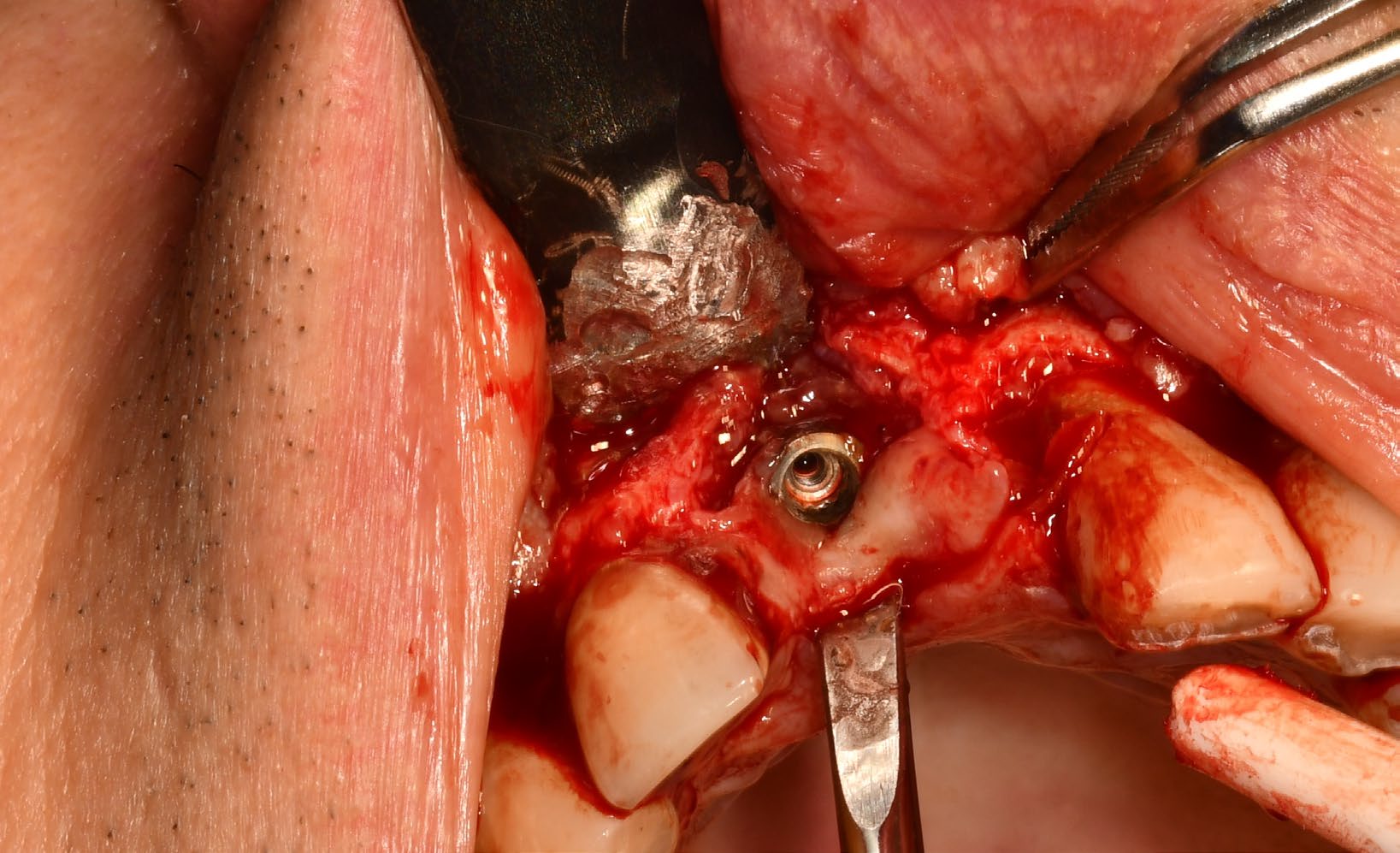
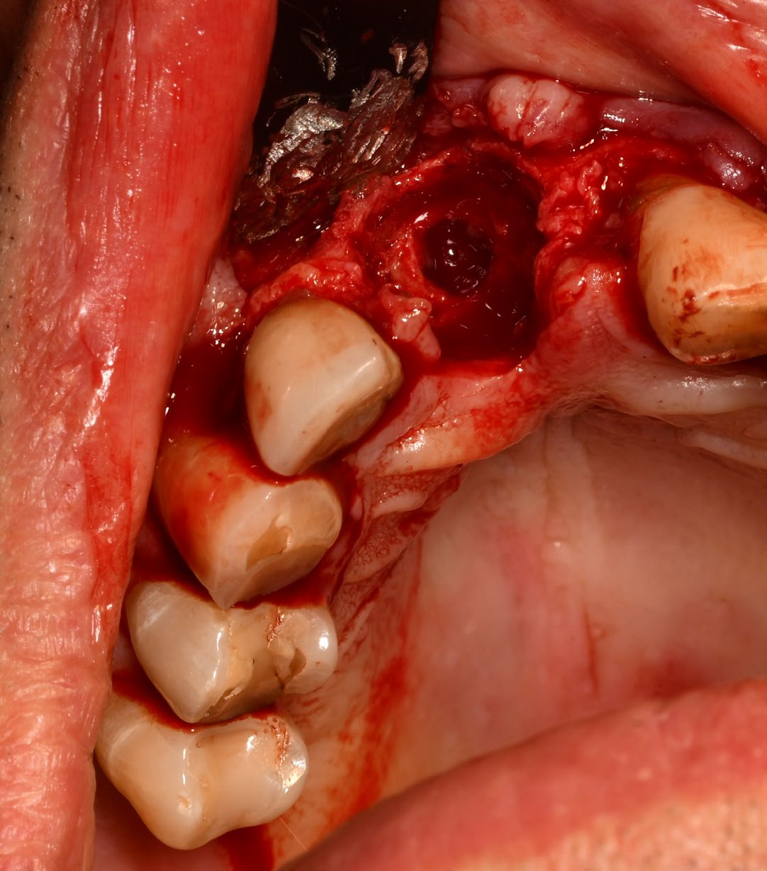
Adults with active, advanced peri-implantitis clinically indicated for implant explantation.
Implant surfaces
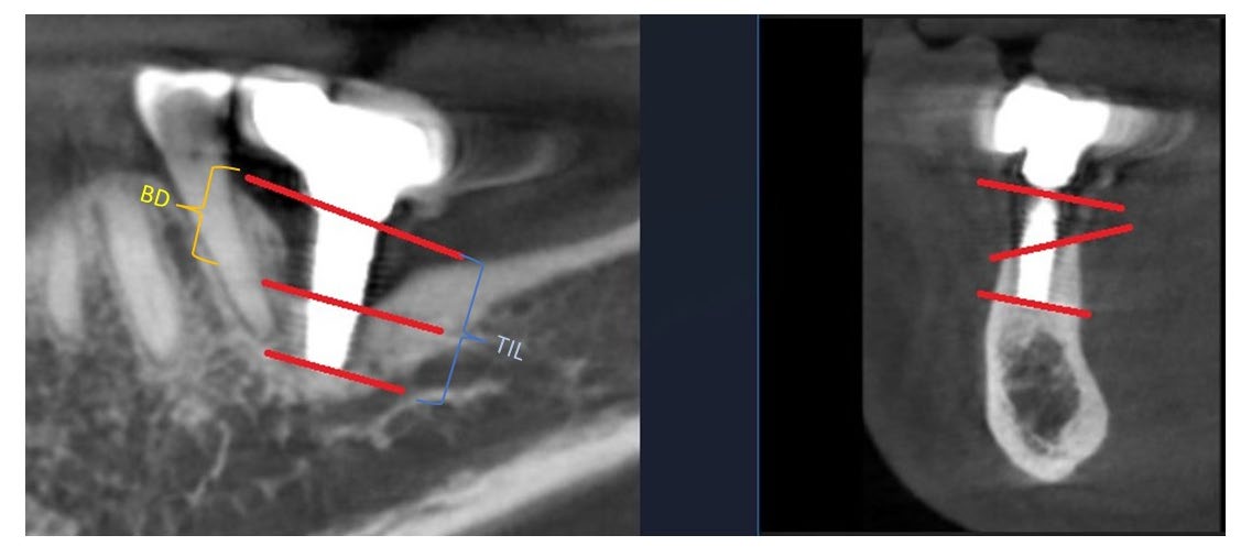
Probing depth and CBCT-based bone loss were assessed at buccal, lingual/palatal, mesial and distal sites.
Follow-up
Peripheral blood was collected before explantation and again four weeks after implant removal.
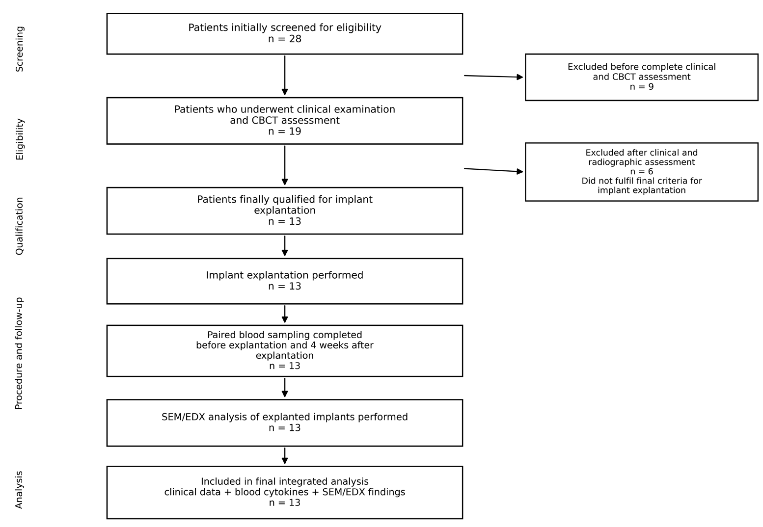
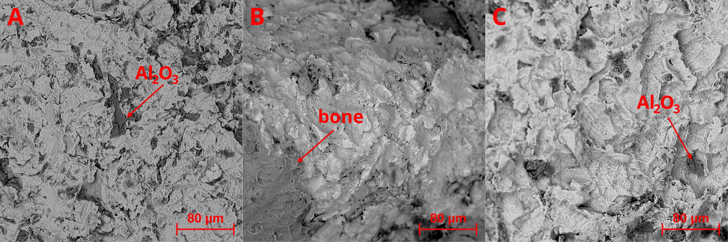
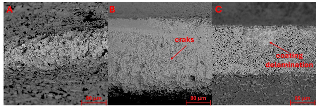
The laboratory panel included IL-1β, IL-6, IL-8, IL-10, IL-12p70, TNF-α, high-sensitivity C-reactive protein (hsCRP) and fibrinogen. After explantation, implant surfaces were evaluated using scanning electron microscopy and energy-dispersive X-ray spectroscopy (SEM/EDX).
Key findings
1. IL-6 tracked the severity of advanced peri-implantitis
IL-6 was strongly associated with both clinical and radiographic severity. Higher circulating IL-6 corresponded to greater mean and maximum probing depth, a larger number of surfaces with probing depth of at least 8 mm, greater mean and maximum bone loss, and more surfaces with at least 80% bone loss. These associations remained significant after correction for multiple testing.
| Severity measure | Association with baseline IL-6 (Spearman rho) |
|---|---|
| Mean probing depth | 0.859 |
| Surfaces with probing depth ≥8 mm | 0.835 |
| Maximum probing depth | 0.831 |
| Surfaces with bone loss ≥80% | 0.823 |
| Mean bone loss | 0.798 |
| Maximum bone loss | 0.742 |
All IL-6 associations shown above remained statistically significant after Benjamini-Hochberg correction. Because this was a small exploratory study, effect estimates require confirmation in larger cohorts.
2. Cytokines did not uniformly decrease after explantation
Four weeks after implant removal, the study did not identify a uniform, statistically significant reduction across the measured cytokines, hsCRP or fibrinogen. This finding suggests that systemic inflammatory activity may persist beyond local surgical treatment or may partly reflect broader host-related inflammatory susceptibility.
3. Corrosion-related findings generated a new hypothesis
SEM/EDX-defined corrosion-related findings were present in five of the thirteen explanted implants. They were not associated with greater baseline clinical severity or higher baseline IL-6 or TNF-α. However, the post-explantation TNF-α pattern differed between exploratory subgroups: the median change was +0.90 pg/mL in the corrosion-related group and −1.16 pg/mL in the group without evident corrosion-related findings. TNF-α increased in four of five patients with corrosion-related findings and decreased in seven of eight patients without such findings.
How should the findings be interpreted?
The results support the view that advanced peri-implantitis may have a measurable systemic immune-inflammatory component. Among the markers studied, IL-6 appears to be a promising indicator of inflammatory burden because it was associated with both soft-tissue findings and radiographic bone destruction. In contrast, hsCRP and fibrinogen did not show the same relationship in this cohort.
The observed TNF-α trajectory in relation to corrosion-related findings is hypothesis-generating rather than conclusive. The study was preliminary, included only 13 patients, had no healthy or disease-control group, and used a short follow-up period. Larger controlled studies with longer observation, paired serum and peri-implant crevicular-fluid measurements, tissue-level immune profiling and quantitative analysis of titanium particles or ions are needed.
Read the full scientific article
Download the complete open-access publication, including detailed methods, statistical analyses, tables, figures and references.
Download the full article (PDF)Explore related PERI-EDU resources
Reference: Dominiak M, Baranowska A, Matys J, et al. Clinical Severity, Pre- and Post-Explantation Cytokine Profile and SEM/EDX-Defined Corrosion-Related Findings in Advanced Peri-Implantitis: Preliminary Results from the PERI-EDU Prospective Exploratory Clinical Study. Journal of Functional Biomaterials. 2026;17:395. https://doi.org/10.3390/jfb17080395