Integrating Peri-Implantitis Research into Higher Education Curriculum
Developing and Integrating Evidence-Based Teaching Materials and Clinical Tools
Research
The PERI-EDU project investigates peri-implantitis as a complex and multifactorial disease developing at the intersection of microbial challenge, host immune response, biomechanical loading, and implant material degradation. Our research goes beyond the traditional plaque-driven model and explores whether peri-implant tissue destruction may also be associated with immuno-osteolytic mechanisms triggered by titanium-derived particles, metal ions, and physicochemical changes on implant surfaces.
To better understand the mechanisms underlying peri-implantitis, the project combines clinical, radiological, biological, and material-based analyses. This interdisciplinary approach allows us to examine both local and systemic factors that may contribute to disease development and progression.
Our research includes the clinical phenotyping of patients with advanced peri-implantitis using standardized parameters such as oral hygiene indices, probing depth, bleeding on probing, peri-implant soft tissue measurements, implant mobility, medical history, and implant- or prosthetic-related variables. These data help us characterize the clinical presentation of the disease and identify factors associated with severe peri-implant tissue breakdown.
An important part of the study is also the radiological assessment of peri-implant bone destruction. Using Cone Beam Computed Tomography (CBCT), we evaluate bone loss around implants on different surfaces and analyse local bone morphology and density. This enables a more precise and objective assessment of disease severity and its relationship to clinical and implant-related factors.
The project further investigates systemic inflammatory and immunological responses associated with peri-implantitis. Blood-based analyses include hematological inflammatory indices, cytokine profiles, acute-phase and nutritional markers, lipid and metabolic parameters, thyroid-related indices, and vitamin D status. By comparing these parameters over time, we aim to determine whether peri-implantitis is associated with a measurable systemic inflammatory signature.
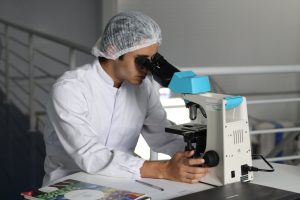
Another major area of research concerns the analysis of explanted implants. Retrieved implants are examined using light microscopy as well as scanning electron microscopy with energy-dispersive X-ray spectroscopy (SEM/EDX). These methods allow us to assess surface morphology, elemental composition, corrosion foci, microstructural changes, and signs of material fatigue such as microcracks.
Special attention is given to corrosion- and fatigue-related mechanisms. We examine whether repetitive mechanical loading and oral environmental conditions may contribute to implant surface degradation, wear, and corrosion. Understanding these processes may help explain why some implants remain stable over time, while others develop progressive peri-implant tissue destruction.
A central objective of the project is the integrated analysis of all collected data. By correlating clinical findings, radiological measurements, inflammatory markers, and material characteristics of retrieved implants, we aim to identify biologically meaningful patterns and improve the understanding of peri-implantitis pathogenesis. This integrative perspective may support future diagnostic, preventive, and therapeutic strategies in implant dentistry.